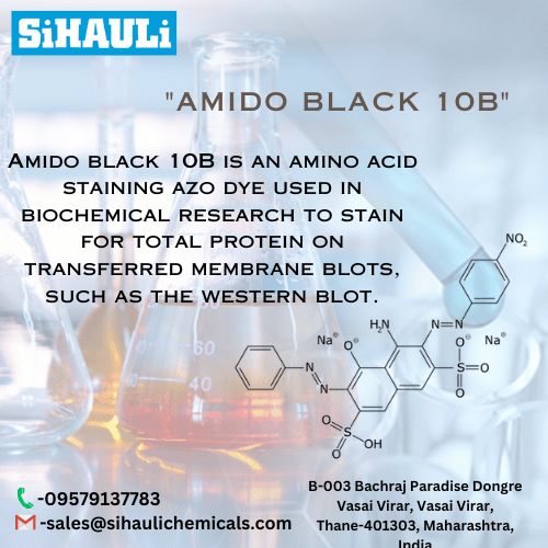
Amido black 10B is an amino acid staining azo dye used in biochemical research to stain for total protein on transferred membrane blots, such as the western blot.
AMIDO BLACK 10B,
IVD In vitro diagnostic medical device
Naphtol blue black, Acid Black 1
For staining protein, collagen, and hemoglobin
INSTRUCTIONS FOR USE
REF Catalogue number: AMB-P-10 AMB-P-25 (25 g)
Introduction
Histology, cytology and other related scientific disciplines study the microscopic anatomy of tissues and cells. In order to achieve a good tissue and
cellular structure, the samples need to be stained in a correct manner. Amido Black 10B is anionic, hydrophilic diazo dye. At first it was used as a
component of Van Gieson trichrome dyes for staining collagen and reticulin, as well as hemoglobin. Today it is also used for quantification of protein
in tissue sections. It may be used as nuclear dye in cytology and histology. In biomedicine it is used for detecting protein, i.e. in milk, as well as for
staining proteins in gel electrophoresis, but also on polymer membranes.
Product description
AMIDO BLACK 10B – powder dye for preparation of solution for staining proteins
Other preparations and reagents used in preparing the solution for staining proteins:
Methanol (CH3OH)
Glacial acetic acid (CH3COOH)
Distilled (demi) water
Preparation of solution for staining proteins (100 mL volume)
Weigh 0.1 g of Amido Black 10B powder dye
Add 40 mL of methanol and 10 mL of glacial acetic acid. Gently mix
Set the volume to 100 mL using distilled (demi) water. Gently mix until all the dye is dissolved.
Keep the solution at room temperature for 6 months. Prepare a new solution after 6 months have passed.
Preparation of solution for destaining (100 mL volume)
Mix 20 ml of methanol with 50 ml of distilled (demi) water.
Add 7.5 ml of glacial acetic acid.
Set the volume to 100 mL using distilled (demi) water. Gently mix
Procedure for staining membrane proteins
- After the proteins have been transferred onto the membrane, rinse the membrane using distilled (demi) water
- Stain using solution for staining proteins 5 min
- Destain using destaining solution 1 – 3 times
- Rinse with distilled (demi) water
Note
The mentioned formulation is only one of the ways of preparing the dye solution. Depending on personal requests and standard laboratory operating
procedures, the dye solution can be prepared according to other protocols.
Preparing the sample and diagnostics
Use only appropriate instruments for collecting and preparing the samples. Process the samples with modern technology and mark them clearly.
Follow the manufacturer’s instructions for handling. In order to avoid mistakes, the staining procedure and diagnostics should only be conducted by
authorized and qualified personnel. Use only microscope according to standards of the medical diagnostic laboratory. In order to avoid an erroneous
result, a positive and negative check is advised before application.
Safety at work and environmental protection
Handle the product in accordance with safety at work and environmental protection guidelines. Used solutions and out of date solutions should be
disposed of as special waste in accordance with national guidelines. Chemicals used in this procedure could pose danger to human health. Tested
tissue specimens are potentially infectious. Necessary safety measures for protecting human health should be taken in accordance with good
laboratory practice. Act in accordance with signs and warnings notices printed on the product’s label, as well as in BioGnost’s material safety data
sheet.


